How does an MRI scan work?
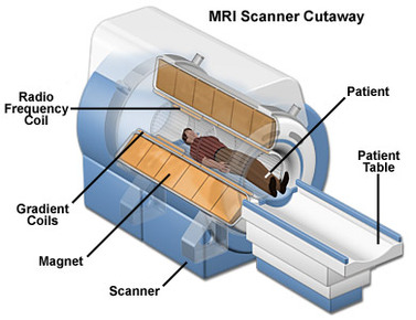
An MRI scanner uses strong magnetic fields and radio waves to create an image of the inside of the body. Hydrogen, oxygen, and carbon atoms compose about 99% of the human body. The magnetic fields and radio waves from the MRI scanner are what affects almost all protons of these atoms, enabling the scanner to create an MRI for almost any part of the body. The radio waves, which are 10,000 to 30,000 times stronger than the magnetic field of the Earth, are sent through the body of the patient during the scan. Both the radio waves and the magnetic field align the protons in the body in one direction. When the magnetic field turns off, these protons return to their original position prior to the application of the magnetic field and radio waves, making the protons send out their own radio waves. The scanner picks up the radio waves of the protons and sends them to a computer, which then develops these radio waves into images of the inside of the body. The images are based on the location and strength of the proton's incoming signals. Different protons send different signals that vary in strength, as it depends on which tissue the proton is found in. For example, a proton from a bone will send a different radio wave signal than a proton found in the blood. This is the reason why an MRI scan can show distinguishable and specific details, therefore medical professionals are able to see different tissue structures clearly, making it easier for them to diagnose their patient accurately.
Preparation and Procedure of an MRI Scan
Before scheduling for an MRI scan, patients should determine if they have any metal or surgical implants that may be affected by the scanners strong magnets. On the day of the patients exam, patients may be recommend by the MRI technologist a certain dietary method that will help in publishing the best quality images. Patients should arrive early for their exam as there may be a copious amount of paperwork. Patients are recommended to wear comfortable clothes that are clear of metal snaps, pins and zippers. If there are issues with the clothing, there are hospital gowns available for the patient. All personal objects such as jewelry, cell phones, keys, and shoes are kept in a locker that the patient is lent for the examination day.
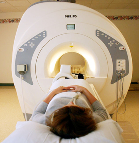
Once the patient enters the exam room, they are assisted by the technologist on the the softly padded bed that gently glides into the scanner. Relaxation is key for MRI scans, and many people are given sedatives to aid in being stationary, which is required for clear images.Technologists ask patients to remain normal breathing patterns and try to decrease anxiety. Patients may also be given injections that will enhance their tissues in the images. Patients are given the option to listen to music and even watch a movie, all things available for the comfort of the patient. The technologist and the patient maintain steady communication during the scan, this is very important. During the scan, loud repetitive noises, banging, clicking, hammering, are heard. When it is silent, the technologist communicates with the patient and prepares them for the next round of noises. The time period of the scan depends solely on the body part that is being scanned. Usual times are 30 mins to 1 hour 30 mins. Laying still is the key aspect of getting an MRI scan done. Scans may have to be repeated if images turn to be blurred and unclear.
After the exam is done, the patient can go back to regular activities, they have no restrictions. The images are sent to the patients doctor for explanations, or the MRI technologist will speak to the patient themselves, and analyze the image and help patients interpret the image.
